
Grafika:Benign gastric ulcer 1.jpg
Z Wikipedii
Grafika w wyższej rozdzielczości jest niedostępna.
Benign_gastric_ulcer_1.jpg (400 × 408 pikseli, rozmiar pliku: 28 kB, typ MIME: image/jpeg)
| | Plik Benign gastric ulcer 1.jpg [ edytuj opis ] umieszczony jest w Wikimedia Commons, repozytorium wolnych zasobów projektów Fundacji Wikimedia. Wyjaśnienie podanej poniżej licencji znajdziesz na stronie Opisy licencji grafiki. |
[edit] Summary
| Description |
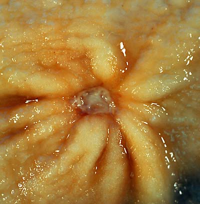
gastric ulcer This 1-cm benign gastric antral ulcer was discovered serendipitously in a gastrectomy specimen removed for adenocarcinoma of the fundus (not shown in the photo). The gross appearance is classic for a benign ulcer in that 1) it is relatively small, 2) the mucosa surrounding the ulcer base does not appear tumefactive, and 3) the radiating rugal folds extend nearly all the way to the margins of the base. Contrast this appearance with that of the malignant gastric ulcer included in this case collection. The criteria for grossly and endoscopically distinguishing benign ulcers from cancer are not absolute, which is why it is necessary to perform a biopsy on any non-healing gastric ulcer. Even biopsies are not 100% accurate in picking up a cancer, so negative pathology reports in such cases may provide false reassurance. The photo was taken with a Minolta X-370 with 100mm bellows lens on Kodak Elite ISO 100 film. The specimen was previously fixed overnight in formalin after being pinned out in a wax-bottomed tray. Photograph by Ed Uthman, MD. Public domain. Posted 23 Sep 00 |
|---|---|
| Source | |
| Date | |
| Author | |
| Permission (Reusing this image) |
PD |
[edit] Licensing
| This file has been (or is hereby) released into the public domain by its author, Ed Uthman. This applies worldwide.
In case this is not legally possible: العربية | Български | Česky | Dansk | Deutsch | Ελληνικά | English | Español | فارسی | Français | Italiano | 日本語 | Nederlands | Polski | Português | Русский | Svenska | Türkçe | Українська | 中文 | 中文(台灣) | +/- |
Historia pliku
Kliknij na datę/czas, aby zobaczyć, jak plik wyglądał w tym czasie.
| Data/czas | Wymiary | Użytkownik | Opis | |
|---|---|---|---|---|
| aktualny | 00:43, 5 cze 2006 | 400×408 (28 kB) | Patho | ({{Information| |Description=gastric ulcer This 1-cm benign gastric antral ulcer was discovered serendipitously in a gastrectomy specimen removed for adenocarcinoma of the fundus (not shown in the photo). The gross appearance is classic for a benign ulcer) |
Odnośniki do pliku
Następujące strony odwołują się do tego pliku:

