Изображение:Epithelial-cells.jpg
Материал из Википедии — свободной энциклопедии

Epithelial-cells.jpg (47 КБ, MIME-тип: image/jpeg)
| | Cведения об этом файле находятся на Викискладе, — хранилище изображений и мультимедиа для использования в проектах фонда Викимедиа. | |
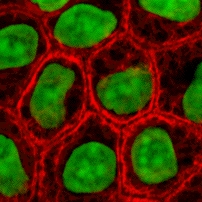
Cultured MDCK en:wikipedia:epithelial cells were stained for en:wikipedia:keratin, desmoplakin, and en:wikipedia:DNA. The stained cells were visualized by scanning laser confocal microscopy. The image shows how keratin cytoskeletal filaments are concentrated around the edge of the cells and merge into the desmoplakin which is located at en:wikipedia:desmosomes of the surface membrane. The network of keratin to desmosome to keratin linking the cells of an epithelial sheet is what holds together tissues like skin.
- This image is taken from the wikibooks Cell Biology textbook (licensed under the GFDL): http://wikibooks.org/wiki/Image:Keratin.jpg
- The copyright to this image is retained by John Schmidt (user:JWSchmidt).
- Permission is granted to copy, distribute and/or modify this image under the terms of the GFDL, as indicated in the fine print at the bottom of this page. If you do not want to use this image under the terms of the GFDL, you can alternatively use it under the terms of the Creative Commons Attribution-NonCommercial-ShareAlike License.
 |
Permission is granted to copy, distribute and/or modify this document under the terms of the GNU Free Documentation License, Version 1.2 or any later version published by the Free Software Foundation; with no Invariant Sections, no Front-Cover Texts, and no Back-Cover Texts. A copy of the license is included in the section entitled "GNU Free Documentation License".
العربية | Česky | Deutsch | English | Español | Français | Italiano | 日本語 | 한국어 | Nederlands | Polski | Português | Slovenčina | Svenska | עברית +/- |
This image can be used under the terms of either the GFDL or the Creative Commons Attribution-NonCommercial-ShareAlike License.
Ссылки
Следующие страницы ссылаются на данный файл:

